The theoretical bases of fluorescence have been known for a long time, and this knowledge allowed the development of myriad cutting-edge technologies, from confocal imaging of cellular processes to super-resolution imaging of single molecules and protein thermodynamics. Yet, despite these advancements, most studies on animal fluorescence are limited to visually stunning photographs taken under controlled lights and filters that not only obscure the underlying mechanism, but also often lead to the erroneous conclusion that fluorescence can make light and thus have an ecological function
One reason for this gap lies in the difficulty of studying fluorescence from organisms as opposed to isolated compounds. Tissues are complex, and fluorescence is just one part of an intricate interplay of multiple structures and pigments that scatter and absorb light. To assess the contribution of fluorescence under real environmental conditions, methods that discriminate between fluorescence and scattering are required.
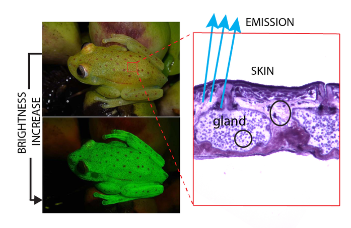
We have already shown that fluorescence can substantially increase the perceived brightness of the treefrog Boana punctata by converting short wavelength light (which many animals are less sensitive to) to longer wavelength light (which many animals are more sensitive to).
We are currently investigating other fluorescent amphibians to elucidate the chemical identity of their fluorophores and to better understand the physical and chemical environments of the tissues that amplify fluorescence emission. We are interested in both visible and near-infrared emission.
In the lab we use a variety of spectroscopic techniques and approaches in photophysics to develop quantitative methods, as well as the theoretical frameworks to model the influence of fluorescence emission under various environmental conditions. We also work in collaboration with visual ecologists to further investigate the visually guided behaviors that are related to fluorescence.
In the lab we use a variety of spectroscopic techniques and approaches in photophysics to develop quantitative methods, as well as the theoretical frameworks to model the influence of fluorescence emission under various environmental conditions. We also work in collaboration with visual ecologists to further investigate the visually guided behaviors that are related to fluorescence.

